Abstract
INTRODUCTION : Immunoglobulin (IGH and IGK) and T cell receptor (TCRG) rearrangements (-R) by polymerase chain reaction (PCR) have emerged as important ancillary tools for assessing clonality of B-cell and T-cell processes, respectively, in specimens such as lymph node, bone marrow and peripheral blood. However, the utility of these gene rearrangement studies in cerebrospinal fluid (CSF) samples has not yet been systematically examined. Cytomorphology and flow cytometry (FC) are currently the standard tools for the assessment of the CSF for leptomeningeal involvement by hematological malignancies. There are inherent limitations to both of these diagnostic methods; thus, it is important to establish the role of IGH/IGK- R and TCRG- R studies in the examination of CSF.
METHODS : Pathology reports of 87 consecutive CSF specimens from 61 patients submitted for IGH, IGK and/or TCRG -R studies in the past three years (2015-2017) were retrospectively reviewed. PCR for IGH (using FR3 and JH primers), IGK and TCRG rearrangements was performed using the BIOMED2 assays. The PCR products for all three reactions were analyzed via polyacrylamide gel or capillary electrophoresis. The interpretation was rendered by an attending molecular pathologist. These results were assessed in the context of the corresponding flow cytometry report.
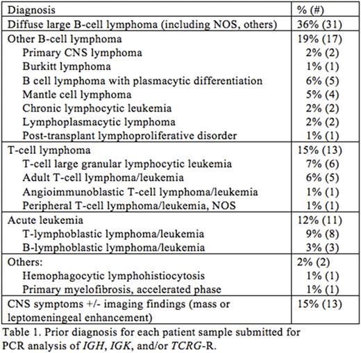
RESULTS : Majority of the patients (82% of patients) had a prior diagnosis of a hematologic malignancy (Table 1), including diffuse large B-cell lymphoma (31% of specimens) and acute leukemias (17%), while the remaining patients presented with CNS symptoms with/without a brain mass. Nearly equal numbers of cases were requested by the clinicians concurrently with flow cytometry (46 cases; 53%) or by the hematopathologist evaluating the CSF FC results (41 cases; 47%). In the latter group, PCR was requested due to an atypical finding based on CSF flow cytometry, which most commonly was an abnormal population with an immunophenotype was different from the diagnostic specimen or a suspicious population that was too small to be diagnostic.
Of the 61 IGH- R and 35 IGK -R tests requested, the majority of the specimens (74% and 63%, respectively) demonstrated a pseudoclonal pattern or poor amplification because of the paucity of B cells. Of the cases with sufficient cellular material, a monoclonal population was detected by IGH- R and IGK -R in more than half of the cases (56% and 54%, respectively). In contrast, in 75% (24/32) of the cases where TCRG-R was requested, there was an adequate number of T-cells for analysis; of these 24 informative cases, 9 (38%) were monoclonal and 15 (62%) were polyclonal.
Overall, 37 of the 87 cases (43%) yielded an informative result for one or more of IGH, IGK, and/or TCRG -R. 38% (14/37) of these cases showed a result that clarified or supported an atypical/suspicious finding by FC, whether submitted by the pathologist (70%) or treating clinician (30%). Eight of the 37 cases (22%) were submitted by the pathologist to confirm the FC findings. In 7 of these 8 cases, the PCR findings corroborated the FC results; there was one discordant case in which a polyclonal T-cell population was seen in a positive FC case of T-lymphoblastic lymphoma/leukemia. Remainder of the cases (14/37, 38%) were submitted by the treating clinicians; if they were submitted together with a concurrent FC analysis (12 of 14 cases), the PCR results confirmed unambiguous FC results (whether negative or positive). In 6 of the 14 cases in which a IGH -R and/or IGK -R identified a monoclonal population, the atypical or neoplastic population constituted <5% of the analyzed cells by FC.
CONCLUSIONS : The paucicellular nature of the CSF specimens provides a challenge not only for cytomorphology and FC but also for PCR for IGH, IGK, and TCRG rearrangements. Nonetheless, an informative clonality result was observed in 43% of the cases submitted for PCR analysis. The results of 60% of these cases (26% of the total submitted cases) were helpful for clarifying and/or supporting an FC finding. Thus, PCR for IGH, IGK and TCRG -R of CSF specimens is a useful ancillary diagnostic aid and can help in the interpretation and confirmation of flow cytometry results, particularly when the neoplastic lymphoid cells are of low abundance or exhibit an immunophenotype that is distinct from the diagnostic tumor.
No relevant conflicts of interest to declare.
Author notes
Asterisk with author names denotes non-ASH members.


This feature is available to Subscribers Only
Sign In or Create an Account Close Modal